There was a lot of heated discussion the past week on Twitter between Seth Trueger (@MDaware), Minh Le Cong (@rfdsdoc), Nicholas Chrimes (@nicholaschrimes) and myself regarding the principles of emergency airway management.
The patient scenario, as I understood it, was someone who was hypoxic to the point of pre-arrest. The crux of the argument was how to secure the airway, whether it is with a supraglottic airway (SGA/LMA) or with endotracheal intubation (ETI). Seth and Minh argued for a first-step SGA insertion after induction, oxygenating the patient through the SGA, and possibly replacing the SGA with an ETI afterwards. Nick and my argument was that an SGA insertion prior to any attempt at laryngoscopy might be an extraneous step that could cause serious problems.
To paraphrase what numerous staff have told me: There is often no right answer, and you can get away with almost anything. But, you must know the benefits, risks, and limitations of each possibility and be able to justify your decision. As long as you can justify the risks and have taken precautions to manage or mitigate the risks, then even if the outcome is bad, you have a leg to stand on in court. With that in mind, I’ll go into some of the reasons why I would have preferred to skip the SGA portion before definitive airway.
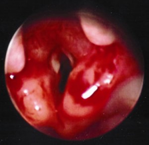
In anesthesia practice, we routinely provide effective bag-mask-ventilation (BMV) with-or-without an oral airway. The very first step of airway management should be a thorough examination of the patient for ease of ventilation and intubation; i.e. “Can I bag that?”. Sometimes you can’t, and you have to use both hands to form a good seal, or use high insufflation pressures (>15mmHg) that could cause insufflation of the stomach, stent open the esophagus and promote emesis/aspiration. When we cannot bag the patient with two hands and accessory airway devices, then we move on to something like an LMA. Though we often use LMAs in routine OR practice in select patients undergoing select operations, it is not a first-line device for BMV. Factors that cause difficult intubation may also cause difficult LMA insertion or seal.
The scenario that Seth and Minh are depicting is a patient who is hypoxic despite being on a non-rebreather or 100% oxygen. The causes of hypoxia are usually listed as 1) low FiO2 2) V/Q mismatch 3) R-to-L shunt 4) diffusion defects 5) severe hypoventilation, and I would also add 6) DO2/VO2 mismatch. There are many clinical etiologies which cause hypoxia, just to name a few: tension pneumothorax, severe pneumonia, aspiration, upper/lower airway obstruction, severe hypovolemia (converts more lung zone 2 to lung zone 1). The crux of my argument for NOT using an SGA initially after induction (additional points to be made later), was that if I could not improve oxygen saturations despite effective BMV synchronized with patient respirations, then a SGA would not contribute much more to those efforts, and in fact, may cause further harm by airway trauma, requiring additional sedation to the patient, or causing emesis/aspiration. Seth and Minh argued that an LMA would help with keeping the patient oxygenated after induction of RSA (rapid sequence airway), buying time before intubation, and that any attempts to intubate the patient would cause immediate life-threatening hypoxia and arrest.
An example of a patient like this could be one with severe bilateral pneumonia. I would add that this patient may be septic, severely hypovolemic, respiratory fatigue, high O2 consumption, severe airway disease and developing ARDS. Their vitals, just for argument sake, HR 120, BP 90/50, SpO2 85% on NRB, RR 35. I think this case is difficult enough to not go into them having a difficult airway, actively vomiting, TBI/ICP etc.
An important point which I would like to make in this situation is that the patient has numerous issues that should be addressed at the same time or before airway management. Early broad-spectrum antibiotics, aggressive fluid resuscitation to improve V/Q matching by recruiting Zone 1 to Zone 2 lungs, and ensuring effective O2 delivery are key to optimizing this patient. Knowing that a hypovolemic, hypotensive patient will crash immediately with positive pressure ventilation and mitigating that is critical. It would be wise to have a second person managing hemodynamics while you’re managing the airway. It is absolutely important to make the first airway manipulation your best attempt, and to have a variety of backup plans available and ready to go because the patient will crash quickly. The backup plan should be something that you are very comfortable with, whether that is video laryngoscopy, SGA, bougie, or surgical.
In my approach to such a critically ill patient, I would probably not give much or any medication before airway support. In a hypovolemic patient, the initial volume of distribution of all drugs will be decreased, and so they will be exquisitely sensitive to induction medications. I would likely try to topicalize with lidocaine spray and then testing if the patient will tolerate laryngoscopy by attempting to insert an oral airway to facilitate synchronous BMV. If the patient was already obtunded from hypoxia/hypercarbia, they would not require anything and may not react to laryngoscopy either. However, they will still have intact airway reflexes and close their cords when you try to intubate. The benefit of just using BMV at this stage to preoxygenate is that you can provide 100% O2 without traumatizing the airway and do not need to give any induction medication. The benefit of using a SGA is mainly if you cannot get a good seal with two hands, you may be able to get a better seal with an SGA which are inserted blindly. However, the patient may not tolerate a SGA insertion without sedation, and depending on the SGA, they have a maximum seal pressure above which leaks will occur. Furthermore, SGAs can be tricky to seat properly and can cause bleeding and trauma. Why not make your first airway manipulation your best attempt with the most definitive airway? If you cannot get a good enough view for intubation with topicalization, then back off and you’ve lost nothing.
From personal experiences with tenuous patients, obese previously-labelled difficult intubations who self-extubated, gross aspiration/vomiting (which you never want to try to ventilate with SGA anyway), I have gotten away with giving little to no medication. The most difficult part is probably intubating a spontaneously breathing patient with intact reflexes. You have to time the insertion of the tip of the ETT with their inspiration, and once they are coughing, have the stylet withdrawn ~1/3 and the rest of the tube advanced with their breaths. Of course this is easier said than done, but I’d prefer a spontaneously breathing patient that I can bag than an apneic patient with a SGA, as would be the case in RSA.
I’d love to hear what others have to say about this, and their own personal experiences in such situations, as I’ve never used SGA first to oxygenate a patient pre-intubation without even attempting BMV.