Category Archives: Anesthesiology

Post dural puncture headaches

This last month I was on a regional anesthesia rotation, which at that hospital, was performed by a dedicated anesthesiologist, fellow and resident in a block room.  We had a handful of referrals for post-dural puncture headaches (PDPH) which we assessed for possible epidural blood patch.

First of all, I will refer to this excellent review article on the topic.

It was interesting to go through some of the decision making processes for whether or not to perform epidural blood patching.

Case #1: This patient developed a sudden onset headache with aural fullness, a “whooshing” sound, neck stiffness, and went to the ED.  There, she had a plain CT head which was reported as normal, and then a diagnostic LP was performed which was negative for infection and blood.  The next few days her headache was worse, and a postural component developed.  At that time, she was being considered for blood patching, but a phone call to the patient revealed that she was doing better, and really had not given enough time to see if the PDPH would get better on its own.

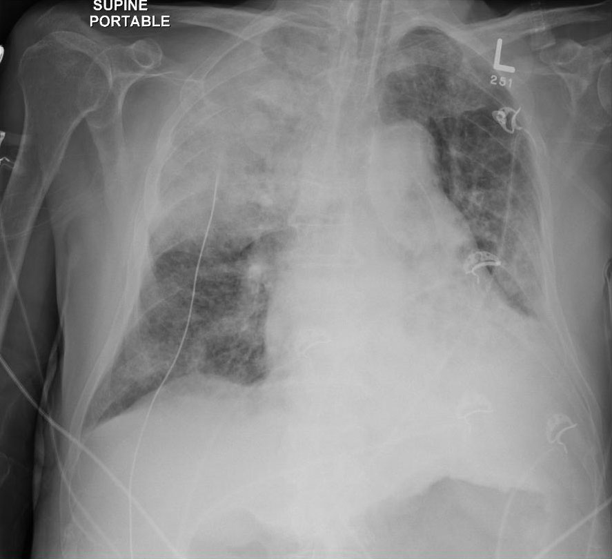
She returned to ED when it got worse and was seen by neurology, who ordered an outpatient MRI.   The MRI a few days latter showed small bilateral subdural collections (could not discern if it was sanguinous), leptomeningeal enhancement and venous sinus engorgement, all signs consistent with intracranial hypotension.  Neurology then recommended the patient undergo blood patching.

When she came to the regional room, she gave a history of still trying to perform at a high level at work, not really taking a break, and having good and bad days.  She was having a good day at the time.  She was still able to go to work for a few hours sometimes, but other times could not drive more than a block.

We had a very lengthy discussion about the risks and success/failure rate of a blood patch.  It was clear that any complication would be unacceptable for such a high functioning individual, and that her expectations of herself were too high given her condition.  We agreed that she would rest more, and we would check up on her to see how she was doing.  If, for instance, the headache was unbearable at 4 weeks, we would then reconsider doing a blood patch, knowing that it is a very invasive procedure with small but very significant risks.

Case #2: This patient underwent intrathecal chemotherapy and developed a classic PDPH 2 days after procedure.  It was so severe that she could not spend much time out of bed, and was quite debilitating.  Upon seeing her in consult, it was felt that she had only had PDPH for 4 days, and that perhaps her conservative management was not optimal in terms of analgesia and caffeine.  We also added an abdominal binder for whatever it’s worth.  To complicate matters and increase her risk with blood patching, she was neutropenic and her CSF was actually negative for malignant cells.  Therefore, we had the risk of seeding cancer into her CSF, and possible infection that would not be detectable in terms of fever/white count at the time of blood patching.

In summary:
– An epidural blood patch is often first suggested to the patient by specialists who do not perform the procedure, and therefore cannot give the patients an informed discussion of risks and benefits.
– It is an invasive procedure with small but significant risks.  If neurological problems of infection occurs, urgent neurosurgery may be required.
– An inadvertent dural puncture with an epidural needle will make the headache and CSF leak worse, and the risk of dural puncture is quoted as being 0.5-1%.  However, all the factors that would make an epidural difficult will increase that risk.
– Conservative management should be optimized and given enough time to self-resolve before resorting to a blood patch, unless there are neurological symptoms or if the headache is extremely severe and debilitating.
– If blood patching fails, it may need to be repeated (maybe several times) and escalating treatments become even more invasive and ineffective.


Hypoxic pre-arrest in critical aortic stenosis – case trouble rounds

You are on call in the middle of a case in OR.  You are paged by CCU to attend to a pre-arrest STAT.  You run up to the CCU, to find the patient on BiPAP, with RT, RNs, the CCU fellow and resident present.  The quick history you get from the fellow is the patient was planned for a TAVI (transcatheter aortic valve implantation) earlier in the week, but was cancelled because he had CHF and was admitted to CCU for diuresis.  About 30min before you were paged, he acutely desaturated to SpO2 65%, ddx was flash pulmonary edema or aspiration.
On arrival at the bedside...

A quick scan of the vitals showed HR 130s AF, NIBP 90/50, SpO2 ~78% with a decent waveform.  Patient was E4 V1 M1, BiPAP on with good seal and oropharyngeal airway in place.  On examination the patient is clearly in resp distress, short shallow breaths.  Legs are grossly edematous, EJ and IJ are visibly distended.  Auscultation reveals a gurgling expiratory sound on right, very decreased air entry on left.  Inspiratory crackles were not obvious.  There was no subcutaneous air or tracheal deviation. The patient had no arterial line, and their only IV was a 22ga antecubital that was attached to a dopamine infusion.

The CCU fellow is suggesting that you give the patient 10-15 mins trial of BiPAP to see if hypoxia will improve.

What are your main concerns with this patient?
1) Airway/Oxygenation – pt is severely hypoxic with unclear etiology.  Hypoxia will worsen myocardial dysfunction and also cerebral ischemia in context of hypoperfusion.

2) Breathing – the work of breathing itself is metabolically high, and taking over respiration with mechanical support will conserve that oxygen for other vital tissues.  He will not improve on BiPAP if the cause of the hypoxia is aspiration.

3) Circulation – remember the main disease is critical aortic stenosis.  The hemodynamic goals with  AS are normal sinus rhythm (maximize LV filling with atrial kick), slow-normal HR (maximize diastolic filling and LV perfusion time), high afterload (need higher myocardial perfusion pressure) and adequate preload (may need higher LAP to fill stiff/hypertrophied LV).  Lack of beat-to-beat invasive BP monitoring, and lack of adequate IV access. Hypoxia and hypercarbia will also increase pulmonary vascular resistance through hypoxic pulmonary vasoconstriction reflexes, and will further impede forward flow.

An arterial blood gas result that was drawn 15 mins ago comes back as pH 7.19 / pCO2 123 / pO2 44 / Bicarb 45 / Base excess 12.

What are the options for the next step?

1) Continue BiPAP – probably not the best option because of aforementioned reasons and hypercarbic/hypoxic respiratory failure

2) Optimize hemodynamics with cardioversion or rate controlling agents – may or may not stay in sinus.  The patient was actually in chronic AF, and thus cardioversion would likely have failed.

3) Intubate.

So you decide to intubate the patient. What is your plan for management of their airway?

I will describe what I chose to do and why.  There may be other safe options, but I chose what I thought would be the safest in my hands.

1) First attempted to perform deep suction with the RT to try to clear out any mucus plugging that may have been causing the decreased air entry I heard on auscultation.  Unfortunately, we did not aspirate much and the patient’s SpO2 did not tolerate lack of PPV very well.

2) I performed manually-assisted BMV in sync with patient’s own respirations as well as intermittent PPV.  The highest SpO2 I could achieve during the ~60s before all my intubation equipment was set up (suction, ETT, scope) was ~76-78%, therefore I decided to proceed.

3) I asked the RN to push phenylephrine 100mcg IV – a total of ~500mcg was given peri-intubation.  Direct look with Mac 3 with no sedation/meds/topicalization.  Rationale was patient was critically ill and already obtunded from hypoxia/hypercarbia.  Gr 2 view with lots of pink froth coming up underneath the epiglottis.  Suctioning did not clear it up – constant stream.  Styleted ETT was placed while patient was spontaneously breathing.  His cords tried to close on the ETT, which was advanced during inspiration, and stylet was removed 1/3-1/2 way and the rest of the ETT was advanced gently through.  Immediately it was filled with pink sputum and this was suctioned with inline suction.

4) We attached the patient to the vent with a starting PEEP of 5 to see if his hemodynamics would tolerate it.  Over the next 5-10mins his SpO2 improved to 88%, which were the highest they’d been in a while.

I stayed for a while longer while the CCU fellow inserted a femoral line and started a dopamine infusion.

Is dopamine a good choice for this patient?

Dopamine was probably not a good choice because of it’s effects on increasing heart rate and pulmonary vascular resistance (PVR).  Pursuant to the SOAP-II trial, norepi might have been a better choice since it increases systemic vascular resistance (SVR) more than dopamine, and does not have such a great effect on HR.  It does, however, also increase PVR.

Vasopressin would probably have been my first-line pressor of choice.  It increases SVR without increasing PVR or HR.  The goal is to improve myocardial perfusion pressure and not overload the RV.

Eventually the patient died later that night on three pressors and after a family discussion regarding futility of further escalation of care.  At the time, I had noticed very deep T waves on the ECG monitor.

This was his preintubation CXR, which was not available to be viewed until later:
preintubation

And this was the postintubation CXR:
postintubation

What I didn’t know until I reviewed the chart afterwards, was that his echo had showed elevated RVSP in the 60s and decreased LV function, both indicative of severe AS and decompensated LV function, causing backup of pressure into the pulmonary vasculature and causing pulmonary hypertension.

Here is a video depicting why tachycardia is so bad for severe aortic stenosis:

Paced tachycardia in critical aortic stenosis from Taylor Zhou on Vimeo.

Take home points:

1) Hypoxia and hypercarbia worsen pulmonary vascular resistance
2) Atrial fibrillation/tachycardia/hypotension bad for aortic stenosis
3) Obtunded, critically ill patients may not need any medications for intubation
4) They will get worse after intubating and initiating PPV before they get better
5) Vasopressin perhaps the best first choice pressor for patients with AS or pulmonary hypertension/RV failure

Emergency airway management

There was a lot of heated discussion the past week on Twitter between Seth Trueger (@MDaware), Minh Le Cong (@rfdsdoc), Nicholas Chrimes (@nicholaschrimes) and myself regarding the principles of emergency airway management.

The patient scenario, as I understood it, was someone who was hypoxic to the point of pre-arrest.  The crux of the argument was how to secure the airway, whether it is with a supraglottic airway (SGA/LMA) or with endotracheal intubation (ETI).  Seth and Minh argued for a first-step SGA insertion after induction, oxygenating the patient through the SGA, and possibly replacing the SGA with an ETI afterwards.  Nick and my argument was that an SGA insertion prior to any attempt at laryngoscopy might be an extraneous step that could cause serious problems.

To paraphrase what numerous staff have told me: There is often no right answer, and you can get away with almost anything.  But, you must know the benefits, risks, and limitations of each possibility and be able to justify your decision.  As long as you can justify the risks and have taken precautions to manage or mitigate the risks, then even if the outcome is bad, you have a leg to stand on in court.  With that in mind, I’ll go into some of the reasons why I would have preferred to skip the SGA portion before definitive airway.

In anesthesia practice, we routinely provide effective bag-mask-ventilation (BMV) with-or-without an oral airway.  The very first step of airway management should be a thorough examination of the patient for ease of ventilation and intubation; i.e. “Can I bag that?”.  Sometimes you can’t, and you have to use both hands to form a good seal, or use high insufflation pressures (>15mmHg) that could cause insufflation of the stomach, stent open the esophagus and promote emesis/aspiration.  When we cannot bag the patient with two hands and accessory airway devices, then we move on to something like an LMA.  Though we often use LMAs in routine OR practice in select patients undergoing select operations, it is not a first-line device for BMV.  Factors that cause difficult intubation may also cause difficult LMA insertion or seal.

The scenario that Seth and Minh are depicting is a patient who is hypoxic despite being on a non-rebreather or 100% oxygen.  The causes of hypoxia are usually listed as 1) low FiO2 2) V/Q mismatch 3) R-to-L shunt 4) diffusion defects 5) severe hypoventilation, and I would also add 6) DO2/VO2 mismatch.  There are many clinical etiologies which cause hypoxia, just to name a few: tension pneumothorax, severe pneumonia, aspiration, upper/lower airway obstruction, severe hypovolemia (converts more lung zone 2 to lung zone 1).  The crux of my argument for NOT using an SGA initially after induction (additional points to be made later), was that if I could not improve oxygen saturations despite effective BMV synchronized with patient respirations, then a SGA would not contribute much more to those efforts, and in fact, may cause further harm by airway trauma, requiring additional sedation to the patient, or causing emesis/aspiration.  Seth and Minh argued that an LMA would help with keeping the patient oxygenated after induction of RSA (rapid sequence airway), buying time before intubation, and that any attempts to intubate the patient would cause immediate life-threatening hypoxia and arrest.

An example of a patient like this could be one with severe bilateral pneumonia.  I would add that this patient may be septic, severely hypovolemic, respiratory fatigue, high O2 consumption, severe airway disease and developing ARDS.  Their vitals, just for argument sake, HR 120, BP 90/50, SpO2 85% on NRB, RR 35.  I think this case is difficult enough to not go into them having a difficult airway, actively vomiting, TBI/ICP etc.

An important point which I would like to make in this situation is that the patient has numerous issues that should be addressed at the same time or before airway management.  Early broad-spectrum antibiotics, aggressive fluid resuscitation to improve V/Q matching by recruiting Zone 1 to Zone 2 lungs, and ensuring effective O2 delivery are key to optimizing this patient.  Knowing that a hypovolemic, hypotensive patient will crash immediately with positive pressure ventilation and mitigating that is critical.  It would be wise to have a second person managing hemodynamics while you’re managing the airway.  It is absolutely important to make the first airway manipulation your best attempt, and to have a variety of backup plans available and ready to go because the patient will crash quickly.  The backup plan should be something that you are very comfortable with, whether that is video laryngoscopy, SGA, bougie, or surgical.

In my approach to such a critically ill patient, I would probably not give much or any medication before airway support.  In a hypovolemic patient, the initial volume of distribution of all drugs will be decreased, and so they will be exquisitely sensitive to induction medications.  I would likely try to topicalize with lidocaine spray and then testing if the patient will tolerate laryngoscopy by attempting to insert an oral airway to facilitate synchronous BMV.  If the patient was already obtunded from hypoxia/hypercarbia, they would not require anything and may not react to laryngoscopy either. However, they will still have intact airway reflexes and close their cords when you try to intubate.  The benefit of just using BMV at this stage to preoxygenate is that you can provide 100% O2 without traumatizing the airway and do not need to give any induction medication.  The benefit of using a SGA is mainly if you cannot get a good seal with two hands, you may be able to get a better seal with an SGA which are inserted blindly.  However, the patient may not tolerate a SGA insertion without sedation, and depending on the SGA, they have a maximum seal pressure above which leaks will occur.  Furthermore, SGAs can be tricky to seat properly and can cause bleeding and trauma.  Why not make your first airway manipulation your best attempt with the most definitive airway?  If you cannot get a good enough view for intubation with topicalization, then back off and you’ve lost nothing.

From personal experiences with tenuous patients, obese previously-labelled difficult intubations who self-extubated, gross aspiration/vomiting (which you never want to try to ventilate with SGA anyway), I have gotten away with giving little to no medication.  The most difficult part is probably intubating a spontaneously breathing patient with intact reflexes.  You have to time the insertion of the tip of the ETT with their inspiration, and once they are coughing, have the stylet withdrawn ~1/3 and the rest of the tube advanced with their breaths.  Of course this is easier said than done, but I’d prefer a spontaneously breathing patient that I can bag than an apneic patient with a SGA, as would be the case in RSA.

I’d love to hear what others have to say about this, and their own personal experiences in such situations, as I’ve never used SGA first to oxygenate a patient pre-intubation without even attempting BMV.

Dr. Levitan’s tips for video laryngoscopy

Supraglottic edema and blood

Dr. Levitan recently posted an excellent article on four tips to video laryngoscopy.  Here are my summary and thoughts:

  1. Suctioning and epiglottoscopy:
    A clean, clear pathway is crucial for successful video/indirect laryngoscopy.  Blood/vomitus/copious secretions will complicate indirect laryngoscopy immensely.  For elective/semi-elective situations, a antisialologue (e.g. glycopyrrolate 0.4-0.8mg IV) can be used, if tachycardia can be tolerated.  For emergency situations, very bloody airways or profuse, active vomiting, two working suctions are key.
    The first step is to identify recognizable structures such as the epiglottis, especially when airway anatomy may be very different in difficult airways.  Even with direct laryngoscopy, find the epiglottis first and then move the blade tip into the vallecula.  As long as you can identify the epiglottis, even if you cannot identify a normal glottic opening, you can try to pass a bougie in the general direction under the epiglottis.
  2. Lift the mandible to expand the viewing space:
    I find the benefit with using Glidescope vs. direct is using less force lifting the jaw.  The curve of the blade itself is sometimes enough to create room to see the glottis, and sometimes lifting can obscure your view or pull the glottic apparatus away from the centre of the screen, thus artificially making the glottis more “anterior”.  When passing the Glidescope blade, just follow the curve of the blade and try not to do any lifting at first.
  3. Tilt the optics away from the target (toward the ET tube):
    The main point is to try to get the glottic opening in the upper 1/2 to 1/3 of the screen, to allow visualization of your ETT approaching the arytenoids.
  4. Two stage tube delivery:
    Dr. Levitan’s suggestion of “STOP, POP, and DROP” is essentially the way that I intubate spontaneously breathing patients who often get “laryngospasm” when trying to insert the ETT through the cords.  Vocal cord closure is to be expected in a spontaneously breathing patient with intact airway reflexes, but this can cause significant problems for users who are not familiar intubating this way.  The key point to getting the tube through is to time the advancement with the patient’s inspiration, since this will open the cords.  If the patient is too obtunded and cannot control their airway reflexes, they may go into full laryngospasm and attempt to breathe against a closed glottis (which can cause negative pressure pulmonary edema).  A light sedated or purely topicalized patient should not do this.  Once the tip of the ETT is in, the cords will close down around it and the patient will start to cough.  This is contraindicated if the patient was actively vomiting / raised ICP / or any other reason why they should not cough or buck the tube (e.g. globe rupture).  At this point, disengage the stylet partly and advance the ETT.
    Another tip for possible “anterior larynxes” on Glidescope is to add a very acute curve on the stylet.  This may cause the ETT tip to go anterior to the glottic opening, but upon partial withdrawal of the stylet, the natural curve of the ETT will bend posteriorly.

This was a very good article from Dr. Levitan which helps to highlight some of the common obstacles encountered with indirect laryngoscopy and how to troubleshoot them.  A frequent complaint I hear from emerg or medicine residents on ICU is that their senior or supervising person steps in too quickly if they run into trouble, and they don’t get the opportunity to troubleshoot themselves.  I still believe a certain level of proficiency with ‘normal’ airways in controlled situations is necessary before attempting to troubleshoot in difficult situations (I include ICU, traumas, and any emergency airway in which there is little preparation time or ability to examine the airway).

Dr. Levitan is an emergency medicine physician, and also founder of Airway Cam Technologies Inc. Image credit: Lurie Children’s Hospital.